THE MOVING BODY - Walking Gait Analysis (Part II): Gait goals and functional anatomy
INTRODUCTION
In part one of this series of articles on the walking gait, we aimed to cover how our gait is constructed and answer the question: What happens during walking? While doing that, we highlighted the main points of interest and stepping stones along the full walk cycle.
In this post, we will try to take those basics and go more in-depth, looking at the gait from a slightly more functional and anatomical angle. We will cover the main muscle groups, timings, and bone structures responsible for moving us forward and answer the question: What makes a walking gait successful?
This might require some previous knowledge of how muscles work and the basics of the walking gait cycle. If you haven't read them, I invite you to stop at part one of this series and this series of short posts. (Walking Gait Analysis for Animation, Part I: https://www.animatornotebook.com/learn/walking-part-1, Anatomy in Motion series part I: https://www.animatornotebook.com/blog/anatomy-in-motion-1)
To cover those articles briefly:
At any given point, at least three forces are acting on the joints: body weight (or gravity), ligament tension, and muscular activity. Generally, we take advantage of gravity where we can to gain momentum and save energy, we use our muscles to counter the force of gravity where needed, and we take advantage of the elastic property of our tendons and muscles to save energy during cyclical motion, building up tension to release it on the opposite swing direction.
We can classify three types of muscle contraction in the body that are generally used for different goals during the walking gait cycle.
Concentric muscle contraction happens the moment a muscle contracts and shortens. This action is usually found in places where the limb is open-chained and needs to be moved somewhere else, for example, while swinging the leg.
Eccentric muscle contraction occurs while the muscle is contracting but lengthening at the same time. Eccentric action is usually found in moments where our body needs to resist external forces or decelerate. In the case of the gait, that could be fighting gravity to keep the body upright.
Isometric contraction happens when a muscle is contracting, but no movement is happening. Isometric contraction generally produces stability in the body, where the robustness of bones alone might not suffice.
With the basics behind us, this article will be divided into smaller chapters, each containing one of the requirements we need for a successful walk.
To clear a full step, there are three main challenges that the body needs to overcome. These will be the phases we divide the gait into in this article:
- Weight acceptance
- Single limb support
- Leg advancement
While there is some overlap between the tasks and the phases of gait, these three phases occur at different timings during the cycle of the leg. So the part covered will tend to focus on a specific moment during the walk.
As always, we will focus our attention on only one leg and side of the body, imagining that in a healthy individual, the other side will be mirrored.
WEIGHT ACCEPTANCE
Weight acceptance happens for the most part in the time between initial contact and continuing during the loading response.
(reference from the right leg)
Weight acceptance takes a larger and more complex role in our bipedal walking cycle. Compared to our quadruped friends, bipeds’ weight is distributed on only two limbs, so they carry a lot more responsibility in resisting gravity at each step we take. Together with our legs, compression on our vertically stacked spine disks also takes a bigger toll than it would if it were lying more horizontally.
The goals of weight acceptance can be divided into two main tasks. First, it is to accept the forward-falling weight of the body, resist gravity, and avoid collapsing to the ground. Second, it is to adapt to the surface the foot is anchoring on. Floors can be irregular, and it is the job of our system to keep the body moving forward without falling off balance on any slightly inclined, twisted, or in any way irregular surface we step on.
Because of the amount of work that is being done, weight acceptance is considered the phase of greater muscle activity. Muscle activity in this phase is generally eccentric (meaning it works to resist lengthening) and affects all the muscles and joints that originate from the spine, extending down to the hips, knees, ankles, and finally the foot itself. A lot is happening at these points, so to hopefully make sense of it all, we will start from the foot/ankle complex and slowly move upstream.
FOOT AND ANKLE ADAPTABILITY
The foot is the foremost part of the body being in direct contact with the ground. When we look at the foot and the ankle in particular, we find several systems that need to be flexible and adaptable to the terrain while at the same time containing many elements that help with shock absorption. Of all the structures we find in the foot, we can divide most of them into two major groups: a passive (mostly comprising the bone structures and fat pads) and an active one (generally muscle tension). Let's break down some of those.
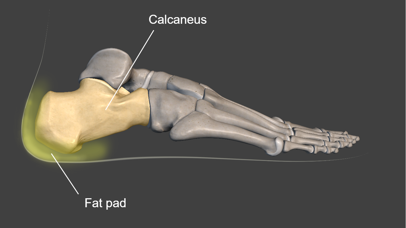
On the passive group, starting posteriorly, we begin cushioning the body from the heel strike and the calcaneal fat pads we find at this extremity of the foot. These pads provide the first impact cushion at a moment where all the force and weight of the body would hit an otherwise narrow surface and would put a lot of stress on the calcaneal bone.
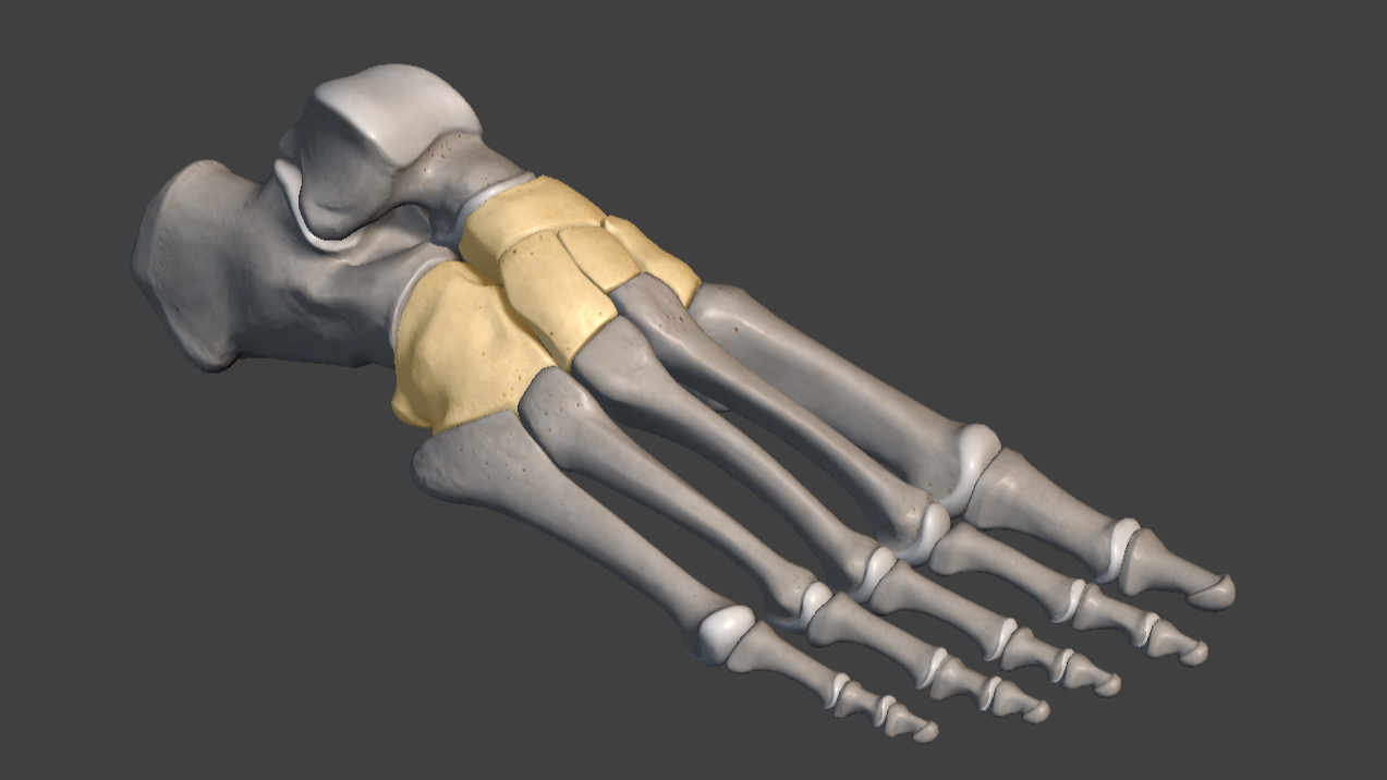
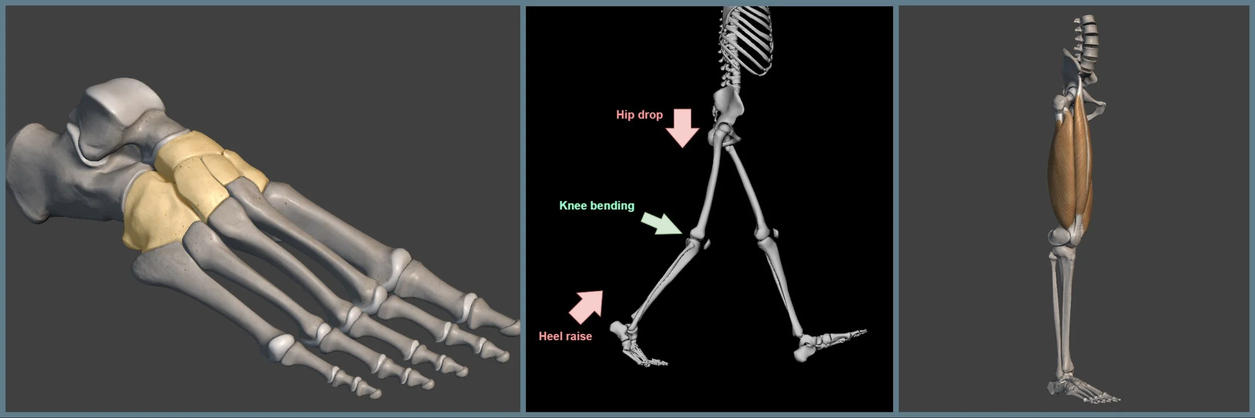
Cuneonavicular joint
Moving slightly anteriorly, and next on the list, we find a series of flat, gliding joints. These types of joints generally are not the most mobile, but they allow for some drifting (as the name "gliding" suggests). This flexibility spreads slightly across the joints on contact to absorb some of the weight of the body. On top of that, the small gliding brings a layer of adaptability to the sometimes uneven terrain the foot is stepping on.

Metatarsal bones
Next, we find probably the most important of the passive subsystems. The metatarsal bones that form the arch characterize the midfoot. The metatarsal bones can spread widthwise and squash to diffuse and neutralize our body weight. At the same time, the arch dome those bones form can controllably "collapse" under pressure to absorb even more of the shock.
An example of how the foot squash, side and top view
A series of ligaments connect all these mentioned joints, adding another layer of reinforcement and shock absorption to the whole structure by binding the bones together like elastic ropes, maintaining some needed flexibility.
Actively, while there are a lot of small muscles present and active in the foot, we will mostly consider two major muscles at work, contracting as the foot touches the ground between initial contact and loading response:
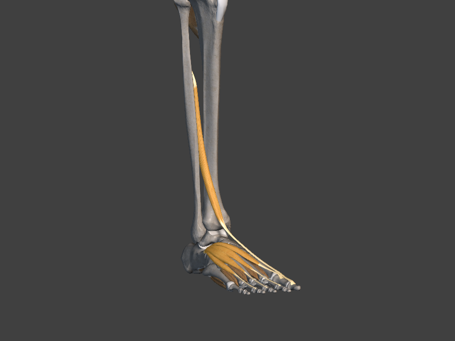
Extensor digitorum
Extensor digitorum: connects from just under the knee to the top part of the toes. It is the muscle generally responsible for bending our toes upward.
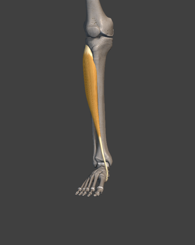
Tibialis anterior
Tibialis anterior: connecting from the lower leg to the inner side of the arch of the foot, this muscle is especially important as it helps support the arch of the foot on contact with the floor, helping to maintain a better alignment of the whole chain of joints above.
Both these muscles start contracting eccentrically slightly before impact, and together they significantly slow down the impact of the foot, preventing it from free-falling and slapping the floor uncontrollably. Instead, we gently release and roll the foot from the back to the front in a slow, controlled fashion.
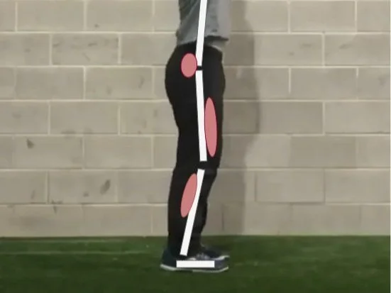
KNEE AND HIP RESTRAINT
The knee and hip joints connect to some of the largest and most powerful muscles in the body. So it's not surprising when we find that the main weight absorption happens at this level.
Even if it's classified as a hinge joint (and hence you would expect it to close and open only along one axis), the bones of the knee actually retain some flexibility to be pushed side to side a few degrees. This flexibility, although small, supports the foot and ankle by absorbing some of the terrain imperfections, helping maintain the rest of the body above in a more straight position.

This side flexibility is then kept safe from injuries by a series of ligaments and small muscles contracting when needed to avoid overextending. However, the main event during the stage of weight acceptance happens along the main axis through the quadriceps.

Quadriceps muscles
The quadriceps are a bundle of four muscles with similar purposes that almost always act together with one common goal: extending the knee and straightening the leg. During the motion we are analyzing, the leg doesn't actually extend, but the quadriceps are nonetheless very active in eccentrically contracting to restrain the amount of knee flexion.
As our weight falls on one of our legs during gait, the natural tendency of the knee joint (together with the rest of the body's skeleton) would be to fold and fully collapse to the ground. At the knee level, the quadriceps are the muscles responsible for keeping the leg straight. During this phase, the muscles also slightly stretch and build up elastic energy that would be used in the phase of push-off.
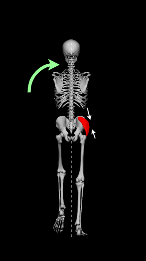
A second layer of muscle resistance to gravity happens around the hips, where the even larger muscles of the glutes must resist the pull downward and at the same time stabilize the hip joint (being a ball-and-socket joint makes it more susceptible to unforeseen movements along all axes).
Because we tend to place our feet towards the midline of the body when we walk, the knee often finds itself not fully aligned with the rest of the leg. Part of the responsibility of the hip-level muscles is to make sure that the knee does not collapse inward, which, over time, can cause imbalances and stress on the joint. Most of the hip muscles are connected to the upper leg on the outside of the bone and, in a balanced body, tension in these muscles rotates the tibia outward and stabilizes the knee, particularly on strides where knee flexion is greater and alignment becomes more important.
Glutes generally tend to activate slightly later than the quadriceps and find their peak contraction in the next phase of gait. But similar to the quadriceps, the hip joint slightly flexes on the loading response, and the job of the glutes is to prevent collapse. At the same time they lengthen, they gather the elastic energy for the push-off that will happen later in the gait.
The glute muscles, together with other muscles located around the hip joint, also act as a trunk stabilizer. By attaching to the lower part of the trunk, they help lift the upper part of the body to avoid folding forward.
Together with it, muscles climb up the spine like the erector spinae, activating to keep the spine straight, counteracting the tendency of the torso to bend towards the ground.
You could, in theory, feel this muscle contract on each step during the walk by putting your hand on the lower part of your back, just on top of the hips.
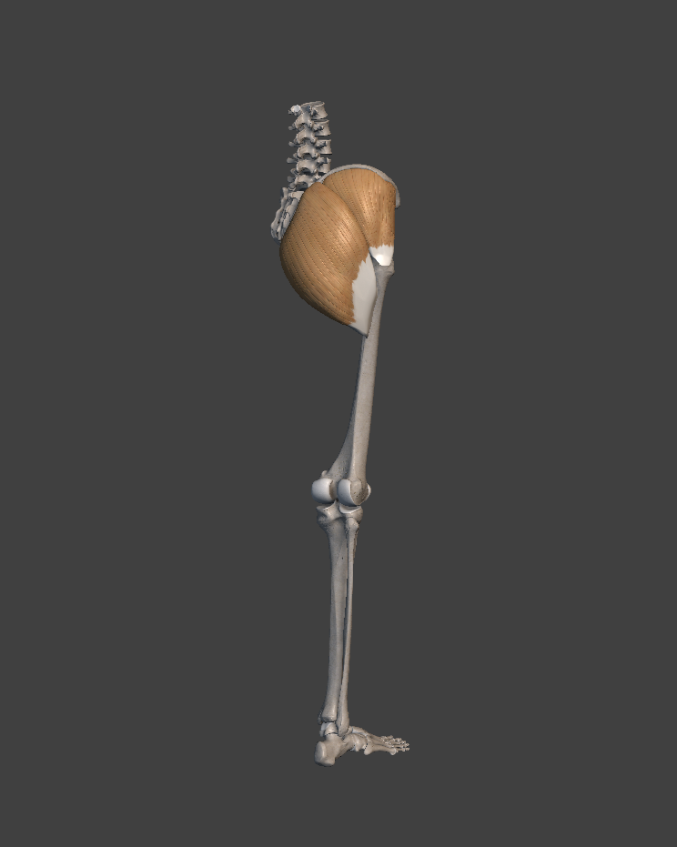
Glutes muscles

Erector Spinae muscles
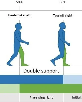
SINGLE LIMB SUPPORT
The main objective of single-leg support is to hold your body weight on a single leg without severe compensation to allow the opposite leg to swing forward. Easy to see, this phase covers the main part of the gait where the opposing foot is lifted from the ground until it reaches forward for contact. Technically, it covers from the loading response to the moment of toe-off, slightly overlapping with the previous phase of weight acceptance.
During this period, the body is transitioning from force absorption to force generation and pushing forward.
We can divide the phase of single limb support into two main parts. The first part covers the motion up until the foot meets the midline of the body, where the body remains in free fall using gravity to advance its position. Here, the main task of support is to catch the weight. The second part of this phase starts from the moment the foot meets the midline of the body, where the leg pushes the ground to drive the body up and forward.
FOOT AND ANKLE PUSH-OFF
While the main shock absorption has happened in the previous phase, the foot doesn't go dormant while holding the body weight on one leg. In the transitional phase before heel-off, the smaller muscles of the foot take the lead. Particularly, the ones curling the toes remain rather active to control our balance and make small adjustments (you can feel this as well while standing still on one foot, your toes should feel quite active).

Soleus and gastrocnemius muscles
During the second part of the heel raise, the bigger muscles connecting to the ankle start becoming more active. The soleus and the gastrocnemius muscle, which are usually seen together as they offer a similar service of heel raisers, have been lengthened up until the moment the heel starts to rise. As always, this moment of stretch builds up elastic energy that can be used on the recoil. Because at the moment of heel-off the center of the body is projected forward, raising the heels won't only push the body up, but also project it forward as well.
During this period of push-off, the heel-raising muscles aren't at peak force production as you would have them if you simply raise your heels from standing. Part of the movement that raises the heels in this phase is passive, as the natural transition of the body moving forward and pulling the back of the leg in extension with it.
Small side fact: the gastrocnemius crosses the knee together with the ankle. While contracting, these muscles also assist the hamstrings in bending the knee for the next phase, all in one action.
KNEE AND HIP BALANCE
Most of the effort at the knee during gait is for force absorption. During the support phase, depending on the personal degree of flexion, the knee finds itself more or less straight. Full extension often means less muscle activation, with the bone structure doing a bigger part in sustaining the weight of the body. Nevertheless, during the whole time as the foot transitions from shock absorption to force generation, the knee-extending quadriceps transition from the first part of eccentric contraction and retaining of muscle elastic energy to concentric extension in the second part of push-off. During heel-off, the knee joint flexes significantly, but this sharp movement is mostly passive and not initiated by muscles. As the hips are falling and the heel is lifting, the space between them is reduced and pressure is put on the leg, forcing it to "shorten" and hence the knee to bend.
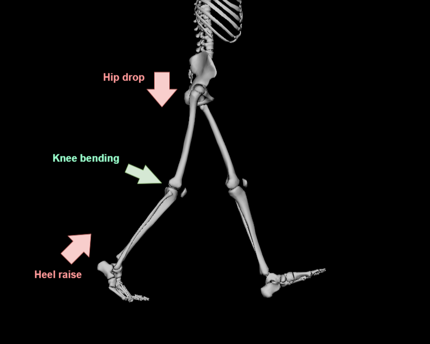
Arguably, the most important and intense effort of single-leg support happens at the level of the hips. The main actors at the hip are the gluteus maximus and gluteus medius. The gluteus maximus is the strongest muscle of the body, and it's used to kick the leg backward or, if the foot is locked on the ground, push our body up and forward. If the quadriceps extend the knees when contracting, the gluteus maximus extends the hip. At the same time, the gluteus medius, which is located more towards the side of the hip, has the very important job of keeping the trunk stable and upright. Because during single limb support our only foot in contact with the ground is not perfectly positioned in the middle axis of the body, the torso has a natural tendency to either twist and bend to maintain balance, or slowly begin to fall towards the non-supported side. The gluteus medius, together with a couple of muscles found on the outside of the leg, keeps the pelvis level. Since the stance phase of the gait can occupy up to 60% of the total gait cycle, a weak or malfunctioning gluteus medius can cause great imbalances and discomfort.

At the level of the torso, the spine muscle's main effort is mostly effective side to side, where it tries to keep the head leveled by compensating for any big or small rotation at the hip. In the gait, the upper body is generally thought of as the passenger, as it usually should remain free to take care of different tasks while the lower body takes us to our destination. Generally, there is significantly little activity on the upper body during a healthy, standard gait. But because our body structures are stacked on top of each other, if there is a weakness or imbalance in the lower body, the torso and the rest of the upper body will have to work harder to compensate and keep us standing.
LEG ADVANCEMENT
We've come now to the last challenge of our gait cycle that will take us from where we left off back to the very beginning, where it all started. Until now, the body's main tasks have been overlapping, and they can be summarized with the words "resist" and "push", as in, we resist the force of gravity, and we push our weight against it. We now find that what is left to do to move forward is quite the opposite; our focus can be summarized as "pull" and "lift".
It is also worth noticing that this phase is the only one where the leg is in an open chain and not restricted. In a closed chain movement, we usually have a point that works as a form of anchor, and the whole body moves around it. For example, if I extend my leg while my foot is on the ground, my foot will not move, and it will be my whole body that will be pushed upward. On the contrary, if I lift my leg in the air and hold it now in an open chain, the act of extending the leg will move my foot further from me while my body remains relatively still. This distinction can be important because although the action of the muscles is the same, we get two very different outcomes.
The phase of leg advancement covers the lifted limb from the moment of toe-off to the end of terminal swing, finishing just at the moment of contact. The main tasks of this phase are two: pull the foot from the back position to the front one, where it can start a new cycle, and lift enough of the foot to clear the floor while transferring it.
FOOT AND ANKLE
At the beginning of this phase, we find the foot and ankle after the last toe was lifted from the ground with some of the residual motion and energy from extending the foot and pushing the floor behind. As the muscles responsible for pushing relax, the foot keeps plantar flexing, and the toes keep curling with it. This motion quickly reverses from plantar flexion to dorsiflexion as the tibialis anterior and extensor digitorum activate and the foot moves towards mid-swing. Contrary to what might be believed intuitively, the foot itself is not being dragged passively towards the front. The tibialis anterior, the muscle that is responsible for flexing the ankle, is working with the extensors of the toes, and together they concentrically contract to lift the foot and toes just high enough that they clear the floor (on average by about one centimeter). With such a small margin, it is no surprise that even in healthy individuals, we can all sometimes stumble if there is any unexpected obstruction on the path.


We could achieve the same clearance by simply lifting and bending the knee more. But it is fair to believe that using the bigger and more powerful muscles of the upper leg for the same purpose will cost more energy compared to simply raising the bottom part of the foot.
The muscle contraction and extension of the foot and toes are isometrically sustained from the mid-swing to the phase of terminal swing and after, preparing for the contact of the foot with the ground and smoothing the roll of the foot during weight acceptance. With isometric contraction, we mean that even though the muscles are active, they remain active to maintain the ankle’s relatively neutral position, and not to increase the angle. In other words, the dorsiflexors stay active, pulling the foot upward, and hold the relative position of the foot even throughout the next phase so that the foot rolls gently on the floor rather than slapping.

(Toes in terminal swing)
HIP AND KNEE
To start detailing the role of the hip and knee muscles, we need to first break down the phase of leg advancement into 3 different stages.
At first, we find the stage of the initial swing, which is the moment when the foot has just left the ground and is preparing to swing. The main goal at this point is to raise the foot vertically. To accomplish that, the hamstrings need to slightly contract, with the help of the passive bending at the foot we covered, to bend the knee and shorten the length of the leg. At the same time, the hip joint needs to move from about 10 degrees of extension to 15 degrees of flexion. For that, we engage our so-called hip flexors, which are two major muscles connecting the lower part of the spine to the front part of the femur. Both these movements combined raise the leg from the ground and begin the swing forward.
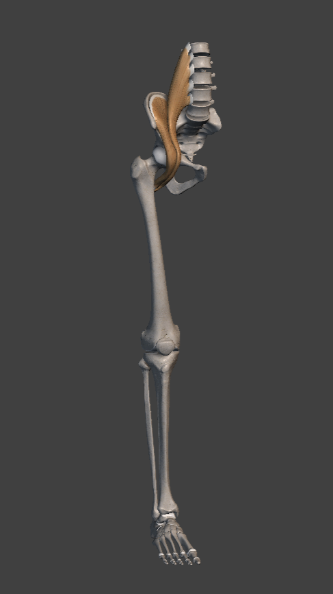
Iliacus and Psoas Muscles
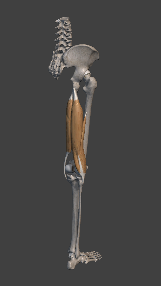
Hamstrings muscles
In the second stage, we are in mid-swing. Here, most of the movement transitions from a bent, backward extending leg towards a front, straightening form. The hamstrings slightly release their contraction to allow the knee to swing forward with the momentum gained and start straightening in front of the body. A small contraction of the hamstrings usually remains present to control the extension. At the same time, our hip flexors contract to bring the leg forward.
The contraction of the hip flexor acting on the swing leg also helps rotate the pelvis forward. On the opposite side of the body, the contraction of the glutes acts in the opposite direction on the stance leg to help stabilize and extend it, rotating the pelvis backward. Taken together, the simultaneous contraction of the hip flexors on one side and the hip extensors (glute muscles) on the opposite side contributes to the rotation of the pelvis at each step and further the reach of the foot.
The third and last stage is the terminal swing, at the end of which we circle all the way back to the start of the whole cycle. As we clear the ground behind us, the only goal remaining is to reach as far as possible. By now, the swing of the leg moving forward is being carried by the momentum created rather than active muscle contraction.
At this point, the body is trying to slow down our joint rotations. At the level of the knee, our hamstring gently contracts to slow down and restrain knee extension. This is particularly important because otherwise, with enough force, the knee could reach into hyperextension and be forced to come to a sudden stop by the hard limits imposed by the knee joint. Pushing our joints to the limit often can, over time, create pain and discomfort, while muscle strain is generally both gentler and more sustainable.
Meanwhile, and on a similar note, the glute in this time frame (maybe slightly anticipating the hamstrings) also activates eccentrically at the level of the hips to slow down and eventually stop the swing forward of the upper part of the leg. Getting ready to contact the ground again.
One last note before ending the cycle. Moments before ground contact, the quadriceps (and some of the muscles necessary for weight acceptance) prematurely begin contracting in anticipation of ground forces to speed up reaction times once the foot touches the floor. Much of what is often needed in a smooth walk is for the body to prepare and anticipate what will happen next. Muscles rarely activate after an event has happened; our brain, through pattern recognition, can anticipate what is needed and prepare accordingly to make sure we are not caught off guard.
WHAT'S NEXT?
Believe it or not, this might not be the end of the story.
In part one, we detailed all the pieces that go into creating a walk, building up from the ground and dividing it into phases to better understand its components.
In this article, we investigated in detail all the intricacies and coordinated efforts of the body; all the actions and timings that the body needs to master to overcome the challenges of something that, from the outside, can look as simple as a successful step.
Now that we understand better what needs to go right during a walk, in the next chapter of this series, we will see some examples of what it looks like when something goes wrong. We will cover some of the more common dysfunctional walks, making sure to detail what they look like, why they look the way they do, and to understand which of the aspects we covered today fail to succeed in making the dysfunction happen.
YOU MAY ALSO ENJOY:
With the knowledge of the previous posts put together, I would like to cover why we tend to anticipate actions. Why does going backward help us move forward, why does going down help us go up, and how does this counterintuitive idea of going in the opposite direction make us move more efficiently?

Bibliography and references
Perry, Jaquelin. Gait Analysis - Normal and Pathological Function.
Rose, Jessica. Gamble, James G. Human Walking.
Palastanga, Nigel. Field, Derek. Soames, Roger. Anatomy and Human Movement.
Vaughan, Christopher L. Davis, Brian L. O’connor, Jeremy C. Dynamics of Human Gait.
Ferber, Reed. Macdonald, Shari. Running Mechanics and Gait Analysis.
Yegian, Andrew K. The Role of Muscles in Arm Swing and Thoracic Rotation During Walking
Desilva, Jeremy. First Steps: How Upright Walking Made Us Human.
Earls, James. Born to Walk.
Arm swing in human locomotion
https://en.wikipedia.org/wiki/Arm_swing_in_human_locomotion
Biomechanics Lecture 11: Gait
https://www.youtube.com/watch?v=QB0tJajDvMw&list=PLS-ocxImwSG_VODKg-Ow12FsyGfDC9cnz&index=11
Observational Gait Analysis - Lecture 1
https://www.youtube.com/watch?v=WsnWnvH3PrM&list=PLwSMRX38xS9AVhulUENkZLCraUn3GM6x3
Observational Gait Analysis - Case Study Review
https://youtu.be/USEtXSRkfAU?list=PLwSMRX38xS9AVhulUENkZLCraUn3GM6x3
THE PHASES OF WALKING (GAIT CYCLE BREAKDOWN)
https://youtu.be/QAnEhz6Eqn4?list=PLwSMRX38xS9AVhulUENkZLCraUn3GM6x3
Phases of the Gait Cycle 1
https://youtu.be/JM0EwSlvR1c?list=PLwSMRX38xS9AVhulUENkZLCraUn3GM6x3
Explaining the Gait Cycle for the NPTE
https://youtu.be/dvpi1WHCDwM?list=PLwSMRX38xS9AVhulUENkZLCraUn3GM6x3
Ground Reaction Force During the Gait Cycle
https://youtu.be/Y2RHvicAM2o?list=PLwSMRX38xS9AVhulUENkZLCraUn3GM6x3
Analysis of Gait Motion Frontal Plane
https://youtu.be/SV0dQnimzrg?list=PLwSMRX38xS9AVhulUENkZLCraUn3GM6x3
Analysis of Gait Motion: Transverse Plane
https://youtu.be/H4wXKqmJac4?list=PLwSMRX38xS9AVhulUENkZLCraUn3GM6x3
The Gait Cycle [Part 1] | Initial Contact (IC) → Midstance (MSt)
https://youtu.be/88juEC9vVyE?list=PLwSMRX38xS9AVhulUENkZLCraUn3GM6x3
The Gait Cycle [Part 2] | Terminal Stance (TSt) → Pre-swing (PSw)
https://youtu.be/6eLbHGJhZvA?list=PLwSMRX38xS9AVhulUENkZLCraUn3GM6x3
The Gait Cycle [Part 3] | Initial Swing (ISw) → Terminal Swing (TSw)
https://youtu.be/BI08Ij8-tNA
Analysis of Gait Motion: Sagittal Plane
https://youtu.be/uh7riepyJl4?list=PLwSMRX38xS9AVhulUENkZLCraUn3GM6x3
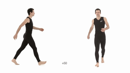
Athletic Male Standard Walk - Realtime. Animation Reference Body Mechanics
https://youtu.be/GBkJY86tZRE?list=PLwSMRX38xS9AVhulUENkZLCraUn3GM6x3
The Gait Cycle
https://youtu.be/-G3EFtkq3qI?list=PLwSMRX38xS9AVhulUENkZLCraUn3GM6x3
Walking Gait Analysis, In Sagital, Frontal and Transverse plane ||Gait Biomechanics
https://youtu.be/OcLFMHTyrnw?list=PLwSMRX38xS9AVhulUENkZLCraUn3GM6x3
Athletic Male Standard Walk. Animation Reference Body Mechanics
https://youtu.be/vq9A5FD8G5w
Human skeletal changes due to bipedalism
https://en.wikipedia.org/wiki/Human_skeletal_changes_due_to_bipedalism
Step by Step: Evolution of Bipedalism
https://efossils.org/book/step-step-evolution-bipedalism
The Catapult Mechanism: Elastic Recoil of Fascial Tissues
https://fasciatrainingacademy.com/the-catapult-mechanism-elastic-recoil-of-fascial-tissues/
Foot and Ankle Structure and Function
https://www.physio-pedia.com/Foot_and_Ankle_Structure_and_Function
Cuneonavicular joint
https://anatomy.app/encyclopedia/cuneonavicular-joint
Tarsal bones
https://anatomy.app/encyclopedia/tarsal-bones
The Gait Cycle
https://www.physio-pedia.com/The_Gait_Cycle
Muscle Activity During Gait
https://www.physio-pedia.com/Muscle_Activity_During_Gait
A Summary of Ankle Dorsiflexion Muscles
https://www.kevinrootmedical.com/blogs/orthotic-news/a-summary-of-dorsiflexion-at-the-ankle-joint#:~:text=The%20main%20muscles%20contributing%20to,anterior%20(Visible%20Body%202019).
Extensor digitorum longus muscle
https://en.wikipedia.org/wiki/Extensor_digitorum_longus_muscle
See how the muscles work to create ambulation
https://canada.humankinetics.com/blogs/excerpt/see-how-the-muscles-work-to-create-ambulation?utm_
Anatomy for the artists app for the muscles
https://www.photojoiner.net/ for the photo edit
https://www.draw.io/ for the diagram